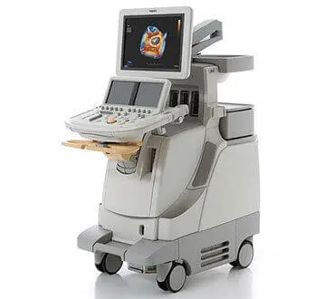
Sonography can be used to examine many parts of the body, such as the abdomen, breasts, female reproductive system, prostate, heart, and blood vessels. Sonography is increasingly being used in the detection and treatment of heart disease, heart attack, and vascular disease that can lead to stroke.
3D Sonography scans show still pictures of your baby in three dimensions. 4D Sonography scans show moving 3D images of your baby, with time being the fourth dimension. It’s natural to be really excited by the prospect of your first scan. But some mums find the standard 2D scans disappointing when all they see is a grey, blurry outline. This is because the scan sees right through your baby, so the photos show her internal organs.
With 3D and 4D scans, you see your baby’s skin rather than her insides. You may see the shape of your baby’s mouth and nose, or be able to spot her yawning or sticking her tongue out. 3D and 4D scans are considered as safe as 2D scans, because the images are made up of sections of two-dimensional images converted into a picture. However, experts do not recommend having 3D or 4D scans purely for a souvenir photo or recording, because it means that you are exposing your baby to more ultrasound than is medically necessary. Some private ultrasounds can be as long as 45 minutes to an hour, which may be longer than recommended safety limits.


