Walking into a specialized clinical space for advanced prenatal screening brings an anxiety that general obstetric visits do not prepare you for. A referral to a specialized fetal medicine and fetal echo center resolves clinical questions about development or cardiac structure instead of providing a standard milestone photo. For patients, transitioning from a standard OB GYN office to a high risk unit feels alarming. For practitioners, this step secures the detailed diagnostic imaging needed to guide management decisions. Understanding how these centers operate, their protocols, and their diagnostic pathways demystifies the process. This environment does not exist to find problems where none exist. It provides clarity, safety, and a precise roadmap for the pregnancy. In practice, most clinics rush patients through these transitions without explaining the shift in tone, which only spikes panic.
The Diagnostic Reality Behind the Referral
Routine prenatal care and specialized fetal assessment differ in their clinical objectives. While a standard obstetric ultrasound checks basic growth, fluid volume, and placental location, a specialized center evaluates organ systems with microscopic scrutiny. Clinicians here use advanced imaging protocols to evaluate structural integrity and physiological function.
Many patients expect a continuation of routine scans, only to find a different pace. The room is quiet, sonographers are intensely focused, and the equipment is more complex. This shift is standard for high level diagnostics. Clinicians map complex anatomical structures to find subtle variations. Every scan targets the specific referral question, meaning the examiner spends significant time on regions of interest like the brain, spine, kidneys, or cardiovascular system.
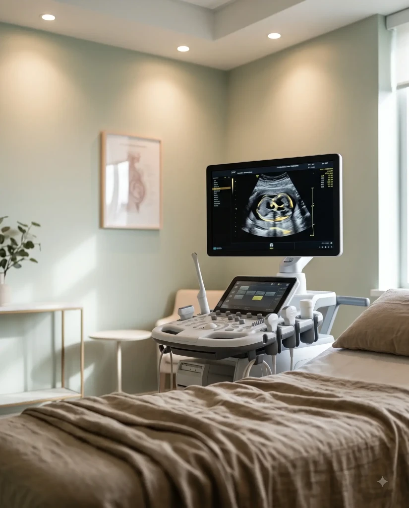
Is a specialized scan different from a routine anatomy scan?
Yes, the difference in scope and depth is substantial. A routine anatomy scan is a screening tool confirming normal development across a broad checklist. If a sonographer notes an atypical finding or if there is a family history of genetic conditions, the patient is referred to specialists. The advanced scan at a dedicated facility is diagnostic. The team actively investigates specific hypotheses, measuring blood flow dynamics through the umbilical cord and brain while analyzing structural symmetry with high definition probes. The clinicians are maternal fetal medicine specialists who completed years of extra fellowship training beyond general obstetrics. They combine ultrasound physics with deep embryology knowledge to interpret findings that a general practitioner might see once in a career. Patients typically receive preliminary information immediately after the scan to reduce the strain of waiting.
Deciphering the Technology at a fetal medicine and fetal echo center
To visualize the womb clearly, specialized facilities use tools far beyond standard 2D imaging. Combining high resolution ultrasound, advanced Doppler, and specialized cardiac software allows clinicians to see structures only millimeters in size. This technology is sensitive to maternal and fetal movement, requiring precise calibration.
The primary tool is the high resolution ultrasound system. Alongside standard imaging, clinicians use color Doppler to map the direction and velocity of blood flow through the fetal heart, brain, and placenta. This detail is vital when evaluating complex conditions like fetal growth restriction or twin to twin transfusion syndrome, where subtle shifts in blood flow dictate delivery timing. Clinicians use 3D and 4D ultrasound technologies to reconstruct complex anatomical anomalies such as cleft lip, skeletal dysplasias, or neural tube defects.
Using these advanced imaging modalities, the diagnostic team creates an accurate, multidimensional map of the fetal anatomy. This allows precise measurement of structures that are otherwise difficult to evaluate, ensuring delivery plans rely on accurate data. These tools provide objective, reproducible measurements to track the progression of specific conditions over time.

Can you do a proper assessment without genetic testing?
While imaging provides a detailed visual map, it cannot read genetic code. Structural variations found on ultrasound are often linked to underlying chromosomal differences. Therefore, imaging and genetic screening are complementary tools. A specialist will often combine a detailed scan with noninvasive prenatal testing or diagnostic procedures like amniocentesis or chorionic villus sampling to get a complete diagnostic picture. Managing a complex pregnancy based on ultrasound alone is like reading a book by only looking at the pictures. You get a sense of the story but miss the precise details needed for postnatal care. Genetic counselors work alongside imaging specialists to help families understand test implications.
Navigating the Patient Experience and Clinical Protocols
Understanding the flow of a typical visit helps reduce anxiety. Unlike a quick checkup, an appointment at a specialized center is a multistep process requiring time, patience, and a collaborative team. Every step maximizes diagnostic accuracy while maintaining a supportive environment.
The visit begins with a medical history review by a nurse or genetic counselor. The imaging session follows, taking forty five to sixty minutes depending on fetal position and case complexity. Patients should prepare for a longer appointment than in general obstetrics because specialists review images in real time and may request additional views. This collaborative review is a standard safety measure ensuring multiple expert eyes evaluate the findings before making a diagnosis. Look, it is not a perfect system and the waiting room delays can be brutal, but having those extra sets of eyes is the most practical safeguard we have.

What happens if the sonographer goes quiet?
When a sonographer stops talking, patients often worry. This silence indicates intense concentration, not a problem. Sonographers perform complex calculations, track rapid fetal movements, and capture precise freeze frames of tiny structures. They are also legally restricted from delivering diagnoses during the scan. The final interpretation must come from the reading physician, who is either a maternal fetal medicine specialist or a pediatric cardiologist. If the room goes quiet, the professional is simply focusing. Patients should write down questions to discuss with the physician during the consultation immediately following the procedure.
The Role of Fetal Echocardiography in Modern Obstetrics
Much of the work at these centers involves fetal echocardiography, a targeted ultrasound evaluation of the fetal heart. Because congenital heart defects are among the most common structural birth variations, precise cardiac imaging before birth is vital. Early identification allows for a coordinated, multidisciplinary approach to delivery and postnatal management.
Prenatal identification of structural heart differences completely changes the trajectory of postnatal care, allowing pediatric cardiologists and surgical teams to be in the delivery room the moment the baby is born.
A fetal echocardiogram is far more detailed than standard cardiac views. The specialist examines the four chambers, outflow tracts, systemic and pulmonary veins, and valve function. This allows early detection of complex conditions like Hypoplastic Left Heart Syndrome, Tetralogy of Fallot, or transposition of the great arteries. Knowing these diagnoses before birth allows families to plan deliveries at tertiary care centers equipped with neonatal intensive care units and pediatric cardiac surgery teams. This preparation improves postnatal outcomes by avoiding emergency transfers and ensuring the baby receives immediate medical intervention.
Why is cardiac screening performed separately?
The fetal heart is a complex, fast moving structure about the size of a peach pit at twenty weeks. Imaging it requires specialized software, high frequency probes, and a skill set that general sonographers do not use daily. A separate cardiac evaluation ensures the examiner focuses exclusively on the hemodynamics and structural integrity of the cardiovascular system without other distractions. This dedicated focus is important for patients with risk factors like pregestational diabetes, IVF conception, family history of congenital heart disease, or atypical findings on routine scans. Separating this scan allows clinicians to perform an exhaustive evaluation of cardiac function, including heart rate and rhythm analysis. In competitive clinical environments, some centers try to bundle these scans to save time, but it consistently leads to missed details.
Securing Expert Care for Peace of Mind
When navigating a complex pregnancy, specialized expertise is invaluable. Our clinical team at the fetal medicine and fetal echo center provides diagnostic accuracy, compassionate counseling, and integrated care planning. If you require a detailed evaluation, a second opinion, or specialized cardiac imaging, please contact our specialists today to schedule a consultation at our fetal echocardiography department, explore our options for high risk pregnancy management, or meet our team of maternal fetal medicine specialists.
Frequently asked questions
At what gestational age is a fetal echocardiogram typically performed?
A fetal echocardiogram is most commonly performed between 18 and 24 weeks of gestation. At this stage, the heart structures are sufficiently developed to be visualized clearly with high resolution ultrasound, though the scan can occur later if new risk factors emerge.
Do I need a doctor’s referral to visit a fetal medicine center?
In most cases, yes. Because these centers provide specialized diagnostic services rather than routine prenatal care, a referral from your primary obstetrician or midwife is typically required. This ensures specialists have access to your medical history before you arrive.
Does a fetal echocardiogram pose any risk to the baby?
No, a fetal echocardiogram is a noninvasive procedure using standard ultrasound technology, which does not involve radiation. Decades of clinical use and scientific research demonstrate that diagnostic ultrasound is safe for both the mother and the fetus when performed by trained professionals.
What should I bring to my appointment at the center?
Bring a copy of your prenatal records, previous ultrasound reports, genetic screening results, a list of current medications, and insurance information. Having these documents ready allows the clinical team to review your history and provide an accurate assessment.
How long does it take to get the results of the scans?
Most specialized centers provide preliminary results during a consultation with the maternal fetal medicine specialist or pediatric cardiologist immediately following the scan. A formal, detailed report is typically sent to your referring obstetrician within twenty four to forty eight hours.